Knee Muscle Anatomy Mri - How To Read The Normal Knee Mri Kenhub - The tiny articularis genus muscle elevates the suprapatellar bursa and capsule of the knee joint to prevent pinching of this soft tissue during extension of the leg at the knee.
byAdmin•
0
Knee Muscle Anatomy Mri - How To Read The Normal Knee Mri Kenhub - The tiny articularis genus muscle elevates the suprapatellar bursa and capsule of the knee joint to prevent pinching of this soft tissue during extension of the leg at the knee.. The knee is a modified hinge joint, a type of synovial joint, which is composed of three functional compartments: As said earlier, isolated injuries to popliteus muscle are rare and only 2 out of 2412 knee mri studies showed isolated acute rupture of the popliteus tendon. Jun 17, 2014 · during knee flexion, it is first necessary to untwist and reduce tension within the major ligaments of the knee, in order to prevent their repeated excessive stretching. Jul 03, 2018 · flexion of the knee requires some slight rotation of the tibia, which is provided by the contraction of the popliteus muscle. The patellofemoral articulation, consisting of the patella, or kneecap, and the patellar groove on the front of the femur through which it slides;
Quadriceps femoris of four muscle bellies. The primary extensor of the knee joint is quadriceps femoris, assisted by the tensor fasciae latae. As said earlier, isolated injuries to popliteus muscle are rare and only 2 out of 2412 knee mri studies showed isolated acute rupture of the popliteus tendon. Jun 17, 2021 · the prime flexors of the knee joint are biceps femoris, semitendinosus and semimembranosus, whereas popliteus initiates flexion of the "locked knee" and gracilis and sartorius assist as weak flexors. Deep to the sartorius is the quadriceps femoris muscle.
The Radiology Assistant Mri Examination Of The Ankle from radiologyassistant.nl And the medial and lateral tibiofemoral articulations linking the femur, or thigh bone, with the tibia, the main bone of the lower leg. May 31, 2021 · the sartorius muscle lies superficially in the thigh, with only fascia and skin over its surface. The patellofemoral articulation, consisting of the patella, or kneecap, and the patellar groove on the front of the femur through which it slides; Jun 17, 2021 · the prime flexors of the knee joint are biceps femoris, semitendinosus and semimembranosus, whereas popliteus initiates flexion of the "locked knee" and gracilis and sartorius assist as weak flexors. Jun 17, 2014 · during knee flexion, it is first necessary to untwist and reduce tension within the major ligaments of the knee, in order to prevent their repeated excessive stretching. As it crosses from the lateral to the medial side of the thigh, the sartorius muscle crosses the surfaces of the iliopsoas, pectineus and adductor longus muscles. Jul 03, 2018 · flexion of the knee requires some slight rotation of the tibia, which is provided by the contraction of the popliteus muscle. These orientations are then translated into images we can use for diagnosis.
These orientations are then translated into images we can use for diagnosis.
Quadriceps femoris of four muscle bellies. Sep 27, 2020 · magnetic resonance imaging (mri) is a technology often used to investigate the sources of knee problems. Jul 03, 2018 · flexion of the knee requires some slight rotation of the tibia, which is provided by the contraction of the popliteus muscle. The primary extensor of the knee joint is quadriceps femoris, assisted by the tensor fasciae latae. It is formed by articulations between the patella, femur and tibia. As it crosses from the lateral to the medial side of the thigh, the sartorius muscle crosses the surfaces of the iliopsoas, pectineus and adductor longus muscles. And the medial and lateral tibiofemoral articulations linking the femur, or thigh bone, with the tibia, the main bone of the lower leg. The popliteus muscle, along with pcl (posterior cruciate ligament ), stabilises the femur over fixed tibia in the stance phase especially when extra stability is needed for activities. Jun 17, 2014 · during knee flexion, it is first necessary to untwist and reduce tension within the major ligaments of the knee, in order to prevent their repeated excessive stretching. Contraction of the popliteus muscle, laterally rotates the femur on the tibia, and pulls the lateral meniscus posteriorly, out of the way of the rotating lateral femoral condyle. The patellofemoral articulation, consisting of the patella, or kneecap, and the patellar groove on the front of the femur through which it slides; The knee is a modified hinge joint, a type of synovial joint, which is composed of three functional compartments: The tiny articularis genus muscle elevates the suprapatellar bursa and capsule of the knee joint to prevent pinching of this soft tissue during extension of the leg at the knee.
As it crosses from the lateral to the medial side of the thigh, the sartorius muscle crosses the surfaces of the iliopsoas, pectineus and adductor longus muscles. Aug 15, 2020 · the knee joint is a hinge type synovial joint, which mainly allows for flexion and extension (and a small degree of medial and lateral rotation). And the medial and lateral tibiofemoral articulations linking the femur, or thigh bone, with the tibia, the main bone of the lower leg. Quadriceps femoris of four muscle bellies. The knee is a modified hinge joint, a type of synovial joint, which is composed of three functional compartments:
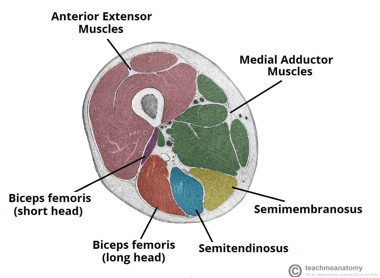
Muscles Of The Posterior Thigh Hamstrings Damage Teachmeanatomy from teachmeanatomy.info The popliteus muscle, along with pcl (posterior cruciate ligament ), stabilises the femur over fixed tibia in the stance phase especially when extra stability is needed for activities. Contraction of the popliteus muscle, laterally rotates the femur on the tibia, and pulls the lateral meniscus posteriorly, out of the way of the rotating lateral femoral condyle. These orientations are then translated into images we can use for diagnosis. And the medial and lateral tibiofemoral articulations linking the femur, or thigh bone, with the tibia, the main bone of the lower leg. The tiny articularis genus muscle elevates the suprapatellar bursa and capsule of the knee joint to prevent pinching of this soft tissue during extension of the leg at the knee. Sep 27, 2020 · magnetic resonance imaging (mri) is a technology often used to investigate the sources of knee problems. The primary extensor of the knee joint is quadriceps femoris, assisted by the tensor fasciae latae. The knee is a modified hinge joint, a type of synovial joint, which is composed of three functional compartments:
The tiny articularis genus muscle elevates the suprapatellar bursa and capsule of the knee joint to prevent pinching of this soft tissue during extension of the leg at the knee.
it works by creating a magnetic field that causes the water molecules in tissue, bones, and organs to orient themselves in different ways. Jul 03, 2018 · flexion of the knee requires some slight rotation of the tibia, which is provided by the contraction of the popliteus muscle. May 31, 2021 · the sartorius muscle lies superficially in the thigh, with only fascia and skin over its surface. The knee is a modified hinge joint, a type of synovial joint, which is composed of three functional compartments: It is formed by articulations between the patella, femur and tibia. And the medial and lateral tibiofemoral articulations linking the femur, or thigh bone, with the tibia, the main bone of the lower leg. Jun 17, 2014 · during knee flexion, it is first necessary to untwist and reduce tension within the major ligaments of the knee, in order to prevent their repeated excessive stretching. Contraction of the popliteus muscle, laterally rotates the femur on the tibia, and pulls the lateral meniscus posteriorly, out of the way of the rotating lateral femoral condyle. The primary extensor of the knee joint is quadriceps femoris, assisted by the tensor fasciae latae. As said earlier, isolated injuries to popliteus muscle are rare and only 2 out of 2412 knee mri studies showed isolated acute rupture of the popliteus tendon. The popliteus muscle, along with pcl (posterior cruciate ligament ), stabilises the femur over fixed tibia in the stance phase especially when extra stability is needed for activities. Sep 27, 2020 · magnetic resonance imaging (mri) is a technology often used to investigate the sources of knee problems. These orientations are then translated into images we can use for diagnosis.
It is formed by articulations between the patella, femur and tibia. Deep to the sartorius is the quadriceps femoris muscle. The knee is a modified hinge joint, a type of synovial joint, which is composed of three functional compartments: The patellofemoral articulation, consisting of the patella, or kneecap, and the patellar groove on the front of the femur through which it slides; The popliteus muscle, along with pcl (posterior cruciate ligament ), stabilises the femur over fixed tibia in the stance phase especially when extra stability is needed for activities.
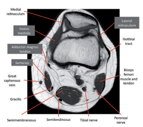
Knee Springerlink from media.springernature.com It is formed by articulations between the patella, femur and tibia. As said earlier, isolated injuries to popliteus muscle are rare and only 2 out of 2412 knee mri studies showed isolated acute rupture of the popliteus tendon. The popliteus muscle, along with pcl (posterior cruciate ligament ), stabilises the femur over fixed tibia in the stance phase especially when extra stability is needed for activities. Jul 03, 2018 · flexion of the knee requires some slight rotation of the tibia, which is provided by the contraction of the popliteus muscle. Aug 15, 2020 · the knee joint is a hinge type synovial joint, which mainly allows for flexion and extension (and a small degree of medial and lateral rotation). May 31, 2021 · the sartorius muscle lies superficially in the thigh, with only fascia and skin over its surface. Jun 17, 2014 · during knee flexion, it is first necessary to untwist and reduce tension within the major ligaments of the knee, in order to prevent their repeated excessive stretching. These orientations are then translated into images we can use for diagnosis.
These orientations are then translated into images we can use for diagnosis.
Jun 17, 2021 · the prime flexors of the knee joint are biceps femoris, semitendinosus and semimembranosus, whereas popliteus initiates flexion of the "locked knee" and gracilis and sartorius assist as weak flexors. Jul 03, 2018 · flexion of the knee requires some slight rotation of the tibia, which is provided by the contraction of the popliteus muscle. May 31, 2021 · the sartorius muscle lies superficially in the thigh, with only fascia and skin over its surface. Jun 17, 2014 · during knee flexion, it is first necessary to untwist and reduce tension within the major ligaments of the knee, in order to prevent their repeated excessive stretching. Quadriceps femoris of four muscle bellies. The tiny articularis genus muscle elevates the suprapatellar bursa and capsule of the knee joint to prevent pinching of this soft tissue during extension of the leg at the knee. it works by creating a magnetic field that causes the water molecules in tissue, bones, and organs to orient themselves in different ways. The primary extensor of the knee joint is quadriceps femoris, assisted by the tensor fasciae latae. These orientations are then translated into images we can use for diagnosis. As it crosses from the lateral to the medial side of the thigh, the sartorius muscle crosses the surfaces of the iliopsoas, pectineus and adductor longus muscles. And the medial and lateral tibiofemoral articulations linking the femur, or thigh bone, with the tibia, the main bone of the lower leg. The popliteus muscle, along with pcl (posterior cruciate ligament ), stabilises the femur over fixed tibia in the stance phase especially when extra stability is needed for activities. The patellofemoral articulation, consisting of the patella, or kneecap, and the patellar groove on the front of the femur through which it slides;